Accidentally Discovered, X-Rays are Essential: From Medicine to Food Production.
Have you ever wondered how X-rays came to be? Though taking an X-ray at the dentists’ might seem normal nowadays, see-through vision had been unheard of until recently in history.
Wilhelm Conrad Roentgen, a German physicist, actually discovered X-rays by accident in 1895. He did so when noticing a glow coming from a chemically coated screen while testing whether cathode rays (now known as electron beams) could pass through glass. Observing how these glowing rays of electrons impacted the glass and their unknown behavior, Rontgen called them “X-rays.” Later, he discovered that X-rays could go through human flesh but not high-density objects like bone or lead (1).
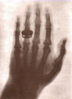
One of Roentgen’s most famous experiments late in 1895 was utilizing X-rays to photograph the hand of his wife, Bertha. This discovery was a breakthrough and became well received in the scientific community (2). Since Roentgen had been using standard cathode tubes, many scientists were able to replicate his experiment. Not long after Roentgen’s discovery, potential applications of X-rays in medicine and surgery became a reality. Several medical radiographs were constructed in the United States and Europe, which surgeons used to guide their work. Just 6 months later, battlefield physicians started using X-rays to find bullets in wounded soldiers (1).
While X-rays had immense potential in almost any scientific field, they were limited to medicine with little industrial use prior to 1912; the source of X-rays, X-ray tubes, broke down under the necessary voltages to penetrate larger objects needed for industrial purposes. These tubes provide the vacuum necessary so electrons fired don’t hit air molecules, as well as a metal target that converts their energy into radiation used for imaging. In 1913, high vacuum X-ray tubes designed by William Coolidge, a chemist and engineer at General Electric, introduced a new, intense, and reliable X-ray source, operating at up to 100,000 volts. A few years later, in 1922, a 200,000 volt X-ray Tube was invented, enabling radiographs of thick steel components. At a seemingly exponential pace, in 1931, the General Electric Company developed the 1,000,000 volt X-ray generator, an extremely effective tool for industrial radiography (1).
With the rapidly increasing use of X-rays, health risks became more evident, especially in the late 19th century. In the beginning, experimenters and physicians continued their work with X-rays without concern about potential dangers (2). In that time period, without much experience with the new technology, nothing had suggested these rays were dangerous. While we now know these rays can be extremely destructive towards the human body, these scientists never thought that a ray similar to light—unseen, unfelt, otherwise invisible—would be harmful for humans.
Tragically, the widespread reckless use of X-rays led to injuries and deaths, including loss of limbs and cancer. X-rays emit high-energy radiation that shatter DNA strands within human cells, leading to genetic mutations and tumors. However, these side effects were often not attributed to X-rays because of the slow symptoms and lack of evidence (1). As time went on, it became obvious that X-rays were at play. They are a form of electromagnetic radiation similar to light, but with a shorter wavelength. This extremely short wavelength gives X-rays the power to penetrate materials that normal light cannot (3). However, if the material is a living tissue, these rays may alter the structure and function of cells, leading to deformities and cancer. Unfortunately, much of this information was discovered from personal experience early on.

In the present day, X-rays continue to be prevalent in daily life. X-rays are still used for medical, industrial, and photographic use. Today, however, high-quality images can be produced, and the process has become automated digitally (3). The usage of X-rays has also revolutionized security systems at airports, welding and casting, and even the production of canned foods, through inspection. So, whether you are watching your bag pass through security or simply opening a can of soup, remember the century of innovation behind that moment. What all started as an accidental discovery is now a necessity that runs the world.
Works Cited
- Nondestructive Evaluation Techniques : Radiography. (2026). Nde-Ed.org. https://www.nde-ed.org/NDETechniques/Radiography/index.xhtml
- Versant Physics. (2023, April 21). X-Ray Evolution: An Accident that Revolutionized Healthcare. Versant Medical Physics and Radiation Safety. https://www.versantphysics.com/2023/04/21/x-ray-evolution-an-accident-that-revolutionized-healthcare/#:~:text=1895,without%20the%20risk%20fully%20understood.
- Stark, G. (1998, July 20). X-ray | Definition, History, & Facts. Encyclopedia Britannica. https://www.britannica.com/science/X-ray/Production-and-detection-of-X-rays
Images
- (2026). Nde-Ed.org. https://www.nde-ed.org/NDETechniques/Radiography/Graphics/Hand.jpg
- Patel, R. B. (2017, February 2). What Is a CT Scan? WebMD. https://www.webmd.com/cancer/what-is-a-ct-scan


Comments are closed.