The science behind immunostaining
Consider a hypothetical scenario where you, someone who needs glasses, do not have them, and you are looking for a pen spring on a dark-colored floor. You can imagine that finding the spring is hard. However, in order to diagnose cancers and develop drugs, biologists have to see proteins within cell and tissue samples (1). To see proteins that are thousands if not millions of times smaller than a pen spring, biologists have utilized a technique called immunostaining since the 1940s. Immunostaining, which derives its name from its reliance on the immune system, utilizes the immune system’s incredible ability to detect small things.
In order to understand the difficulty behind seeing small things, one must understand the concepts of resolution and contrast. The idea of resolution is similar to that of blurriness. The resolution limit of an observer, such as a camera, microscope, or human eye, is the smallest distance in which the observer can distinguish two points as separate objects (2). On the other hand, contrast is the difference in light intensity and color between an object and its background (3). Returning to the hypothetical from the beginning, it is so difficult to find the pen spring because, without your prescription glasses, objects are harder to differentiate, increasing your personal resolution limit increases. Similarly, the pen spring does not contrast with the dark floor, so it blends in and is harder to see.
With the help of electron microscopes, scientists have achieved a resolution limit of approximately 0.1 nanometers, which is several thousand times smaller than the wavelength of visible light (4). An electron microscope’s resolution limit is low enough to observe proteins, but a low-resolution limit is not useful if there is not sufficient contrast between the protein and its background.
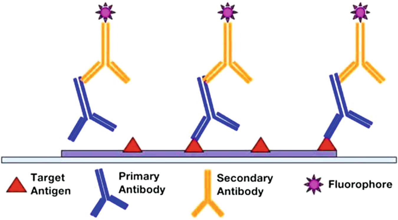
Cells and proteins are mostly transparent, so they do not contrast with the petri dish or slide that they sit on. To give these small objects more contrast, scientists utilize antibodies. When exposed to foreign molecules, also called antigens, the immune system produces a small protein known as an antibody tailored to bind onto the antigen (5). The ability to customize antibodies to bind onto almost any antigen means that biologists can create antigens that bind to a protein they want to see; the antibody that binds to the target protein is the primary antibody. Scientists then produce a secondary antibody that attaches only to the primary antibody. To increase contrast, scientists then attach a dye molecule to the second molecule. Although the dye molecule could simply be attached to the primary antibody, having both a primary and secondary antibody allows biologists more customization as one secondary antibody can bind to many primary antibodies. This also saves on the production cost of antibodies, which are regularly $2,500 for ten microliters.
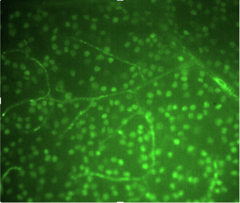
Immunostaining is the process of attaching the primary antibody to the protein and then attaching the secondary antibody to the primary antibody. In hypothetical terms, immunostaining is equivalent to spray-painting your pen spring neon green. The dye molecule attached to the secondary antibody connected to the primary antibody and therefore the target protein provides the sought-after contrast. By using microscopes and immunostaining, biologists now have enough contrast and resolution to observe proteins.
Proteins are everywhere within our body, so being able to detect and observe specific proteins gives biologists a powerful tool to learn more about cells and tissues. Immunostaining is a cornerstone of biology research, and countless world-changing discoveries in the future may utilize this technique.
Bibliography:
- Overview of Immunohistochemistry (IHC). Thermo Fisher Scientific. https://www.thermofisher.com/us/en/home/life-science/protein-biology/protein-biology-learning-center/protein-biology-resource-library/pierce-protein-methods/overview-immunohistochemistry.html#:~:text=Immunohistochemistry%20(IHC)%20combines%20anatomical%2C,their%20target%20antigens%20in%20situ
- Resolution. MicroscopyU. https://www.microscopyu.com/microscopy-basics/resolution
- (2018, September 11th). Contrast in Optical Microscopy. National High Magnetic Field Laboratory. https://micro.magnet.fsu.edu/primer/techniques/contrast.html#:~:text=Contrast%20is%20defined%20as%20the,to%20the%20overall%20background%20intensity
- Electron Microscopy tutorial. The University of Utah. https://advanced-microscopy.utah.edu/education/electron-micro/#:~:text=The%20wavelength%20of%20electrons%20is%20much%20smaller%20than%20that%20of,lens%20system%20in%20electron%20microscopes
- How to make an Antibody. Biolegend. https://www.biolegend.com/en-us/making-antibodies


Comments are closed.