
How three new brain imaging innovations can help scientists better understand neurological disorders
When you think about what the brain looks like, the first image you visualize is probably its rounded, pinkish wrinkled surface; not the 100 billion neurons and 1000 trillion synapses hidden inside (1). However, for scientists researching brain activity and function, having a precise image of its internal neural working is vital for discovering and treating neurological disorders. Specifically, researchers need to study different levels of the brain’s organization simultaneously, from brain cells’ anatomical and physiological architectures to their organ-wide connectivity. By connecting information across multiple scales, scientists can create a comprehensive picture of neural circuits and activity patterns within the brain. However, scientists have historically struggled with capturing such a complex picture (1).
That was until June 2024, when researchers at MIT’s Chung Lab developed and published a technology pipeline that did just this (3). Traditionally, scientists cut each brain hemisphere into incredibly thin slices by a vibratome machine to picture the brain. Then, the slices are stained by dyes with different proteins grabbing onto certain cell types to capture each cellular identity. When scientists scan each slice and form multiple maps of the brain’s cellular population later, sections with a certain protein appear a certain color. So when they come back to analyze their images, they can visually decipher the concentration of each protein. Then, each map is combined and reconstructed to develop a uniform picture of the brain (4). However, this method can take years even for a small chunk of a mouse’s brain, and any mistakes in the process ruin the product (2). Places for error are also common as most vibratomes are not tailored for cutting large brains, distorting some components and making it nearly impossible to realign them into a brain map (5).
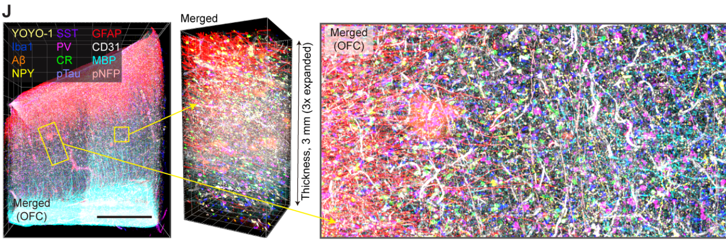
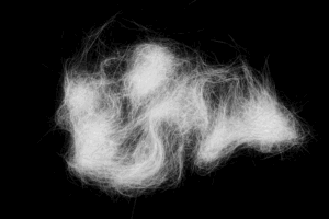
The resulting image of a section of human brain tissue zoomed out. Each color labels various different cells, vasculature and proteins.
Led by Kwanghun Chung, an associate professor at The Picower Institute for Learning and Memory at MIT, a team of researchers developed three new key innovations that upgrade past technologies: MEGAtome, mELAST, and UNSLICE.
The pipeline starts with the MEGAtome. Unlike other vibratomes, the MEGAtome is engineered to vibrate side-to-side at higher frequencies so that slices don’t get damaged or lose anatomical information. Because of its high accuracy in cutting parallel to keep the brain intact, the MEGAtome can cut thicker slices off the brain, shortening the procedural time from months to just days (3).
To assist with the stability of the brain slices, the Chung Lab also developed a hydrogel called mELAST. This transforms and expands brain tissue from previously weak and brittle tissue into a stretchy brain-gel hybrid that can maintain its shape under pressure. In addition, these thicker slices can withstand more rounds of dyes, meaning scientists can capture multiple protein changes in the same tissue at single-cell resolution (6).
The third innovation was the computational system called UNSLICE which recreates the 3D structure of the brain. To use UNSLICE, researchers at the Chung Lab used blood vessels as a guide to roughly align each piece. Then they zoomed in on individual neural connections to perfect the map. This technology allowed scientists to map both the connectivity and the molecular details of individual cells in the human brain. With these innovations combined, the Chung Lab’s upgraded pipeline yields more data in less time (7).
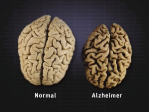
This study does not present an atlas of the entire brain, rather it is a presentation of an integrated suite of technologies that allows scientists to efficiently probe other brain tissues (3). For example, later on, Chung and Mattew Frosh, an Alzheimer’s researcher at Massachusetts General Hospital, compared two brain structures: one from a healthy individual and the other from a person with Alzheimer’s disease. Using the Chung Lab’s imaging techniques and innovations, they located a deficient amount of neurons in the frontal and cerebral cortex (6). This is only one of the many ways the Chung Lab’s brain imaging innovations can be used. Chung says he envisions creating a brain bank of fully imaged brains from different neurological conditions, helping researchers pursue new studies and make more comparisons (6).
A picture of the orbitofrontal cortex of the brain after using MIT’s new pipeline.
Bibliography
- The Picower Institute. “Multiscale, Multiplexed Brain Imaging at MIT.” YouTube, 17 June 2024, www.youtube.com/watch?v=ji0Oba6FYjA&list=PLABvh_IMOr_k3Xez7sTonX6QzuRrcABXj&index=6. Accessed 19 Jan. 2025.
- Fan, Shelly. “This MIT Device Maps the Human Brain with Unprecedented Resolution and Speed.” SingularityHub, 27 June 2024, singularityhub.com/2024/06/27/this-mit-device-maps-the-human-brain-with-unprecedented-resolution-and-speed/. Accessed 19 Jan. 2025.
- Park, Juhyuk, et al. “Integrated Platform for Multiscale Molecular Imaging and Phenotyping of the Human Brain.” Science, vol. 384, no. 6701, 13 June 2024, www.science.org/doi/10.1126/science.adh9979, https://doi.org/10.1126/science.adh9979. Accessed 19 Jan. 2025.
- Mocerino, Maria. “Mind Mapper: MIT’s New Tech Shows Whole Brain Hemispheres in 3D Detail.” Interesting Engineering, 13 June 2024, interestingengineering.com/science/mit-innovation-brain-hemispheres-mapping. Accessed 18 Jan. 2025.
- Singleman, Corinna. “3D Brain Imaging Tool Provides Holistic Analysis down to Subcellular Features.” GEN – Genetic Engineering and Biotechnology News, 13 June 2024, www.genengnews.com/topics/artificial-intelligence/3d-brain-imaging-tool-provides-holistic-analysis-down-to-subcellular-features/. Accessed 19 Jan. 2025.
- Orenstein, David. “Technologies Enable 3D Imaging of Whole Human Brain Hemispheres at Subcellular Resolution.” MIT News | Massachusetts Institute of Technology, June 2024, news.mit.edu/2024/technologies-enable-3d-imaging-whole-human-brain-hemispheres-subcellular-resolution-0617. Accessed 18 Jan. 2025.
- Guan, Webster. “Scalable Subcellular Resolution Mapping of the Human Brain.” Mit.edu, 19 May 2023, dspace.mit.edu/handle/1721.1/151202?show=full, https://hdl.handle.net/1721.1/151202. Accessed 19 Jan. 2025.
Images
- https://www.youtube.com/watch?v=ji0Oba6FYjA
- https://picower.mit.edu/news/technologies-enable-3d-imaging-whole-human-brain-hemispheres-subcellular-resolution


Comments are closed.